So You Want to Be a… Sonographer
| June 18, 2024Good sonographers possess excellent technical skills, eye-hand coordination, and attention to detail

What will I be doing all day?
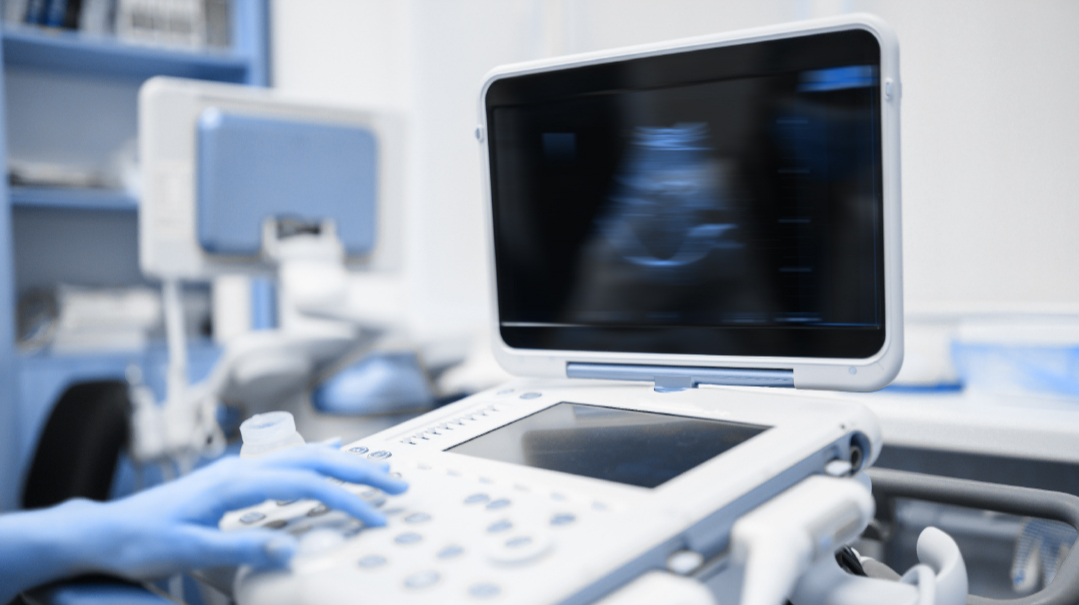
A diagnostic medical sonographer helps physicians diagnose and monitor various medical conditions by using ultrasound imaging machines that produce sound waves inside the body to generate images of internal organs, tissues, or blood flow.
Responsibilities include obtaining patient medical histories, preparing the patient for the ultrasound procedure, operating the equipment, analyzing the images, and collaborating with physicians, radiologists, and other medical professionals to discuss the results.
What kind of career options do I have?
A sonographer can choose to specialize in a range of areas. Common specialties include OB/GYN, abdomen, and superficial structures, echocardiography, vascular technology, musculoskeletal, and neurosonography.
Sonographers work in a variety of health care settings, such as hospitals, private clinics, and diagnostic imaging centers.
What kind of training do I need?
To become a diagnostic medical sonographer, one must complete a two-year associate’s degree program. There are also four-year bachelor’s degree options, as well as a one-year training program for people who already have a degree in the health fields. In addition, the American Registry for Diagnostic Medical Sonography (ARDMS) offers specialized accreditations, which require passing certification exams.
Do I have the personality for it?
Good sonographers possess excellent technical skills, eye-hand coordination, and attention to detail. In addition, they must have good communication and interpersonal skills: patience, compassion, and understanding.
What can I expect to make?
Average salary in the US: $85,000 (salary can vary widely based on location)
TALES FROM THE TRENCHES
TZIPPY FARKAS-BALIN
Brooklyn, New York
Ultrasound Technologist, Lenox Hill Hospital MFM (Maternal Fetal Medicine), New York, New York
Graduated From: B.S. in Radiological Sciences, St. Francis College; DMS (Diagnostic Medical Sonographer), CAHE (Center for Allied Health Education)
Years in Field: 11
My Typical Day at Work
I typically clock in at 9 a.m. at Lenox Hill’s MFM department. I head to my ultrasound room, turn on the machine, and make sure my room is ready for a patient. I then head to the front desk, where all the patients’ charts are (we still have paper charts at Lenox Hill). In our department, whichever technologist is ready takes the next available chart, regardless of what type of exam it is. Some ultrasounds are quick, such as dating exams (performed early in the pregnancy to determinate a fetal heartbeat and how far along a patient is), and second- and third-trimester biophysical profiles, which look at fetal growth and fluid amount and ensure the baby is moving and practicing breathing motions. Other exams take longer and require more precision, such as early or late anatomy scans (usually done around 16 and 21 weeks, where we check to make sure every organ is working and growing properly).
Once I take a chart, I head back to my ultrasound room and review the previous report so I know exactly what is needed for the current sonogram. I then enter the patient’s information into the ultrasound machine, head to the waiting room to call my patient, and bring her and whomever is accompanying her into the room.
I start by introducing myself, tell them what exam we are going to do, and remind them of my role, and that the perinatologist (an OB-GYN who specializes in high-risk pregnancies and fetal ultrasounds) will review the results once we are done. I’ve learned early on to make everyone aware of this prior to starting the exam; if I say it after I start scanning, they automatically assume something is wrong. (Unfortunately, I’ve learned this the hard way.) Also, this way if, chas v’shalom, we do find something wrong, I can simply “remind them” afterward that, as I’ve already said, the doctor will discuss the results, without causing alarm.
Depending on what type of exam we are doing, it can take anywhere between ten minutes to an hour or more, especially if we find something wrong. Once I’m finished scanning, I head out to a computer station where I review my images, to make sure I have what I need and that the images are good enough for the doctor. I type up a report of the visit, including measurements and calculations and anything noted out of the ordinary. I then review the images with the doctor and present my patient and ultrasound findings. Depending on the results, the doctor may scan the patient himself, or tell us to bring the patient to his office where he discusses the findings and answers any questions they may have. I then sanitize the exam table and instruments and make sure the room is properly cleaned for the next patient. On a typical eight-hour shift, we see approximately 12 patients.
Sonographers are also required to make sure each of our individual rooms are fully stocked with appropriate supplies, such as gloves, drape sheets, ultrasound gel, and exam table paper.
How I Chose the Profession
Growing up, I always enjoyed biology class, so when I got older, I wanted to do something in the medical field. I considered becoming a nurse but didn’t love the hours or overnight shifts. When I was in 12th grade, my sister, who was expecting her oldest, suggested I go into sonography. I’ve never looked back.
How I Chose My Specialty
In sonography, there are several specialty options, because there are many different areas of the body you can perform an ultrasound on. When I was in school, I enjoyed echocardiography (ultrasound of the heart) and OB-GYN the most. I did my internship rotation in Lenox Hill’s MFM and upon graduating, the manager reached out to tell me they were looking to hire, and invited me for an interview. I wasn’t ready to look for a job yet, but because this job fell into my lap, straight from Hashem, I knew I had to take it. Today, I use both of my specialty areas in my job; as a maternal fetal medicine sonographer, I also scan the babies’ hearts. I’m lucky to have the best of both worlds.
What I Love Most about the Field
I love being able to interact with people who are experiencing such an exciting period in their lives. I enjoy seeing the excitement on their faces when they get to see their baby. I also feel privileged to be able to help calm patients who are anxious. While we’re limited in what we can say to the patients about what we’re seeing on the screen, just being there to talk and help them relax makes such a difference. Sometimes, talking to the patients and pointing out the baby’s face, hands, or feet can help calm their nerves.
What I Find Most Challenging about the Field
One of the biggest challenges is when you find an abnormality or lack of a fetal heartbeat. Most patients coming to an OB-GYN sonographer are there for a happy occasion. They’re not expecting something to be wrong with their babies. If we find an abnormality, we need to remain composed. It can be quite taxing.
I’ll Never Forget When
Working in Lenox Hill, I get to meet a lot of different kinds of people from all walks of life. We even have some famous celebrities and athletes that come to our practice. I’ve also had the privilege of performing my niece’s anatomy scan.
Something I Wish People Knew about Sonographers
I think, particularly in the field of OB-GYN, people tend to look at our job as being all fun and games. However, we’re not just taking fun pictures of the baby for the parents. Our job is quite serious. We make sure that every organ, every limb, every bone in every finger, and every part of the baby is growing correctly and functioning the way a human body is supposed to be.
How I’ve Seen the Field Change over the Years
A lot of the changes have to do with the machines we use. Thanks to technological advancements, images have become a lot clearer and can focus better on specific areas.
Also, as research develops, there are new images and tests to perform. For example, results of a recent study demonstrated that measuring the waveform of the maternal uterine artery together with the woman’s blood pressure could determine her risk factor of developing preeclampsia (a serious condition in pregnancy that can cause high blood pressure, protein in the urine, swelling, headaches, and blurred vision) as early as 12 weeks into the pregnancy. Thanks to this new information, we can now start an at-risk patient on proper medication and regularly monitor her and her baby.
My Advice for People Starting Out
Be kind to yourself! It can be very discouraging not being able to get the perfect image right away. In order to excel as a sonographer, you need to understand that it takes time, be patient with yourself, and get as much hands-on experience as you can.
Another important attribute as a new sonographer is to be able to accept constructive criticism. There is always something new to discover in this field, and you need to be open to learning and improving.
There currently seems to be a large demand for sonographers. However, many positions do prefer experienced (typically two to three years) technologists, so if you find someone willing to train you straight out of school, grab it. Making good connections is very important and can help you grow and lead you to other job opportunities later on. Personally, I did my rotation in Lenox Hill, got my first job there, left to work somewhere closer to home for a few years, and now I’m back where I started. If I hadn’t left on good terms, or made those strong connections, I probably wouldn’t be there now.
GITTY GELLIS
Ramat Eshkol, Israel
Diagnostic Medical Sonographer, Kupat Cholim Clalit and owner of private home ultrasound business
Graduated From: Downstate Medical Center, Brooklyn, NY RDMS, RDCS (Registered Diagnostic Medical and Cardiac Sonographer), B.S. Certifications in OB-GYN, abdominal, and echo.
Years in Field: 18
My Typical Day at Work
I work part-time in the Clalit medical clinic. (Clalit is one of the four health funds in Israel that provide government-subsidized medical services to every citizen.) My day starts early at 7:30 a.m., for patients coming for fertility treatment, and our ultrasound clinic has a constant stream of patients coming in and out. There are three technicians for the morning shift, and we always help each other out.
A little over two years ago, I started a private home ultrasound business. I bought a top-of-the-line portable ultrasound machine, and patients can choose whether I come to them or they come to my home office, where I have a comfortable ultrasound room set up.
Many of my clients are young newly-marrieds living in Israel, experiencing their first pregnancy. They don’t know what to expect, and maneuvering the Israeli system can be quite overwhelming. There are often extended wait periods to schedule a first ultrasound, with availability only during specific times of day. That’s where my services come in handy. (I’ve even had mothers and mothers-in-law from America call me to schedule appointments for their daughters or daughters-in-law.)
I aim to provide personalized ultrasound services in a calming atmosphere from a warm and experienced American sonographer who speaks their language. My primary goal is to ensure my clients feel comfortable and secure enough to pose any questions or concerns, without them feeling rushed or pressured.
I see all sorts of cases, ranging from ovarian torsion (twisted ovary), which is a medical emergency, to healthy babies, where I simply need to reassure the young parents-to-be that the baby is healthy and everything’s fine.
You have to be flexible for this business; I’m pretty much on call 24/6. I’ve done ultrasounds at the craziest times.
I had ladies come on Friday after the Erev Shabbos siren already sounded (here in Yerushalayim, that’s 40 minutes before shkiah), and at seven in the morning to ensure their babies had heartbeats. Someone came knocking on my door for an ultrasound at midnight on Purim. While ideally clients make appointments in advance, I try to make myself available pretty much any time someone calls.
How I Chose the Profession
I like to work with people and I like the medical field, so I knew I wanted to do something that combined the two. Back when I graduated high school, everyone was going for occupational or speech therapy. I wanted to do something different, and sonography sounded interesting.
I worked at Einstein in the Bronx for three years before moving to Israel 15 years ago.
How I Chose My Specialty
I specialized and took my registry (certification tests) in obstetrics and echo (echocardiogram). I figured whichever area I’d find a job in first is what I’d do — and OB-GYN it was. I enjoy working in the OB-GYN field because people tend to come for happy reasons, as opposed to other ultrasound specialties.
What I Love Most about the Field
When a patient who originally came for fertility treatment ultrasounds comes back expecting. I also love being the one to tell people that they’re having twins (in Israel, the accepted practice is that the sonographer divulges this information); it’s interesting to see the different reactions. And it’s very gratifying when a patient comes in nervous and leaves the ultrasound calm and relaxed.
What I Find Most Challenging about the Field
When I don’t see a heartbeat and have to give over bad news. Also, sometimes in ultrasound, not everything is black and white (no pun intended); we may need to send a patient for a second opinion or tell her to come back for another ultrasound at a later point. At the same time, you don’t want to get a patient nervous unnecessarily.
I’ll Never Forget When
Once, while doing an ultrasound in Clalit, I identified an emergency, and the mother was rushed into the hospital for a C-section ASAP. It’s gratifying to know Hashem put me in a position to save the mother and baby.
Then there were times when the top doctors told the patient it was one gender, and when I scanned, I saw it was the other. (Obviously, it’s not their specialty.)
Something I Wish People Knew About Sonographers
You have to work to get the correct image. Sometimes it’s not as easy as it looks.
Someone sent me a picture of an ultrasound done in America. The newly-trained sonographer had told them the baby was deformed, but in reality, she’d simply taken the image at the wrong angle. Baruch Hashem, the baby was fine; she’d scared the patient unnecessarily.
I only bought my own ultrasound machine after working for 15 years. You have to have a lot of experience before going out on your own.
How I’ve Seen the Field Change over the Years
There are always new things to learn in the field, and I’m constantly educating myself about new developments.
One change I’ve seen here in Israel is that while sonographers used to perform the 20-week anatomy scan, now only doctors do so. (In America, sonographers still perform this scan.)
My Advice for People Starting Out
It can be hard to land your first job, as employers only want to hire sonographers with experience in scanning. If you can’t find a job, offer to do an unpaid internship to get more hands-on practice. Baruch Hashem, I found a job pretty soon after I graduated and learned on the job, but there were people in my program who had a very hard time finding jobs.
It’s ideal to start working in a hospital; even though you’ll get paid less, you’ll be exposed to a wider range of experiences. After getting experience, you can switch to an office.
Also, try to work in a setting where there are other technicians on staff, so that you’ll have someone to ask your questions to (since there are always questions).
RIVKA HELLMANN
Brooklyn,,New York
Diagnostic Medical Sonographer and Program Director and Clinical Associate Professor of Sonography Program, Touro University
B.S. in Diagnostic Medical Imaging, M.S. in Medical Informatics, SUNY Downstate Medical Center. Registry Certifications in Abdomen and Superficial Structures, Obstetrics and Gynecology, Adult Echocardiography, and Vascular Technology
Years in Field: 23
My Typical Day at Work
I have been working in the field of sonography for over two decades and joined the sonography education world 17 years ago.
As a sonographer, a typical day consists of seeing about eight to 12 patients. Depending on what type of sonogram they are coming in for, they may be given preparation instructions such as fasting six hours prior, or drinking four full cups of water and holding it in. By the time the patients see us, they may be understandably irritable. We must calmly and patiently explain the exam, how to situate themselves on the exam table, and give them a gown to change into, if necessary. We triple-check the chart to make sure we have the correct patient and then review the prescription and history to get a clear understanding of why this patient is here and what the doctor needs for us to see in order to make the best possible diagnosis.
Often, patients have no idea why they are coming in for a sonogram other than “my doctor sent me.” That’s when we start asking what I’ve dubbed the Question Quest. “Did you take a blood test or urine test that came back a bit off? Are you in pain? Did the doctor feel a lump or bump somewhere? Do you have a family history of…?”
Some questions get even more personal than that. It’s essential for us to understand why the patient is here, so we can take the best images for a clear diagnosis and help the doctor treat this patient effectively.
The sonogram itself usually takes about 30 to 45 minutes. Afterward, we have the patient wait in the exam room while we review the images with the health care provider on staff that day. Once the doctor is satisfied with the images we’ve acquired, we send the patient home. Some days, we get to assist in special procedures such as biopsies or cyst aspirations. Other days, we struggle getting a wheelchair-bound patient up on the exam table. Every day, we need to put our best foot — and face — forward.
I currently work on the faculty of Touro as the sonography program director and clinical associate professor, a job that involves a wide range of responsibilities and one that I absolutely love. I enjoy collaborating with our wonderful professors to create innovative ways to make our coursework fun and fresh. I also enjoy teaching my students in the classroom setting; their never-ending barrage of question keeps me on my toes. I guide them in practicing different types of sonograms on each other and working with them in our state-of-the-art sonography lab, and I help with clinical placements, track our students’ progress with their clinical supervisor, interview program applicants, and visit local high schools and seminaries in Israel to talk about sonography.
I’m also active in the research side of the sonography field. I’ve published articles in several journals, presented research posters at several sonography and medical conferences, and continue to be involved in research endeavors.
How I Chose the Profession
I always knew I would do something in the “helping” field. Growing up, I was the one who helped my grandfather dress his leg wounds, or took out splinters from my sister’s finger. These things never grossed me out, and I loved the feeling of being able to help relieve others’ pain. At the same time, I was interested in technology, and originally planned on becoming a computer programmer (I actually completed a year of programming at Touro, many years ago). When a neighbor told me about the sonography field, I was intrigued. After a bit of research into schools I was fascinated. I took the year of prerequisites at Touro, got into my first-choice sonography program, and the rest is history.
How I Chose My Specialty
While I’m registered in four specialty fields and am qualified to scan any of those types of sonograms, my “baby,” pun intended, has always been obstetrics and gynecology (OB-GYN) scanning. I still get excited seeing a tiny heartbeat and counting fingers and toes, and love feeling the satisfaction of capturing that perfect baby profile. I love seeing new life begin and being able to see this secret unfold inside the body, while watching the expectant parents’ eager faces — and now the faces of my students performing these sonograms.
What I Love Most About the Field
My favorite part of being a sonographer is the cast of characters I’ve met in the clinical world and now in the classroom. There’s never a dull moment. I also feel so fortunate that the sonographers who train my students at our clinical sites, as well as my faculty and staff, have become my lifelong friends and support system. I feel truly blessed.
I’ll Never Forget When
My most memorable patient was a sweet chassidishe lady who came to me during my first year on the job. It was her tenth pregnancy, and she told me she’d never had a sonogram before, and that all of her previous pregnancies had turned out fine. When her doctor had told her that she was carrying relatively large for the month she was in, she’d waved it off and told him she was just heavy. (Don’t we all say that?) But he persisted and she came to my office.
When I positioned the transducer, I saw this bright white line going through her abdomen and immediately thought it was a dangerous diagnosis. It took me a split second to come to my senses: Tenth pregnancy, large for the stage she was in — duh! Twins! (Remember, this was my first year working.) As we aren’t allowed to share results with patients, I quietly and professionally finished examining and measuring both babies and showed the images to my radiologist. She agreed with my preliminary impression and told me I could tell the patient the exciting news.
I remember being nervous; I wasn’t sure how she would feel about finding out she was having her tenth and now an 11th. But boy, was I surprised — she literally jumped off the exam table and exclaimed, “A tzvilling! A tzvilling!” I didn’t speak Yiddish (sadly, still don’t), but I kind of guessed that “tzvilling” meant twins. She hugged and kissed me and cried tears of joy. Then she skipped down the hall and out the door. Until today, I can still picture the scene in my mind: a short and very pregnant lady literally jumping and clicking her heels as she left the clinic. That was definitely a good day.
Not all stories are happy, though. I remember the first time I had to confirm that a mother had miscarried, six weeks into my job. I cried for a week. There was also a time that someone I knew from my camp days came in for a sonogram during her second pregnancy. Sadly, the baby had so many things wrong that there was no way it was going to survive in utero. The worst part was that I wasn’t even allowed to tell her anything, or offer her a hug. The radiologist told her to call her doctor when she got home. I don’t know what happened afterward, as sonographers often don’t get to know the end of the story.
Something I Wish People Knew about Sonographers
We can see with sound! Sonography does not use radiation like an X-ray; it sends high-frequency sound waves into the body, which bounce off various structures and organs in the body and then bounce back as echoes (pixels). Thousands of pixels form the image we see on the screen. As a sonographer, you become a physics expert in order to really get to know the instrumentation of your machine.
How I’ve Seen the Field Change over the Years
There have been lots of changes. For one, back when I was in sonography school, I remember going out on shidduchim and telling my dates that I was studying sonography. “What? Stenography?” was the inevitable response. No one had ever heard of the field (and the few who had would respond, “Oh, like for the babies?”).
Also, sonographers used to be called “techs” and earned very little respect in the health care field. People simply didn’t understand the importance of our role, the volume of information we need to know, and our value in diagnosing patients quickly, painlessly, efficiently, and non-invasively. Now, everyone knows what sonography is and some folks even go so far as to ask for a sonogram from their doctor. The fact that sonographers got a huge raise across the board in 2023 speaks volumes for how far our field has come and how sonographers have become recognized internationally. And we are finally called “sonographers” and not “techs.”
Each sonographic specialty has also changed significantly over the past 20 years. Some examples: There are new applications in vascular and OB-GYN sonography. Contrast sonography has been introduced to abdominal sonography. Pediatric and musculoskeletal ultrasound registry examinations have now been developed and implemented.
Doctors used to take biopsies “blind,” taking educated guesses as to where to shoot the biopsy gun to sample a piece of tissue. Now, ultrasound-guided procedures have become the norm, with doctors relying on a sonographer to visualize the correct biopsy sample site.
Handheld “butterfly” probes and transducers have been created for paramedics or doctors on the go.
And there are now dedicated sonography machines in the emergency rooms instead of waiting for an on-call sonographer to get to the ER. The list goes on and on. Sonography is an ever-growing technologic field — and we’re just getting started.
My Advice for People Starting Out
While I, personally, for reasons out of my control, have never worked in a hospital setting, I strongly recommend working in a hospital when first starting out. You learn a lot from the doctors in the hospital, the imaging fellows, other sonographers in your department and your patients. Patients you see in the hospital are generally sicker than those who walk in to an imaging facility for an appointment, so you get exposed to many more interesting cases and pathologies. That is definitely the best way to learn.
(Originally featured in Mishpacha, Issue 1016)
Oops! We could not locate your form.


